Our recent paper in Laser & Photonics Reviews presents an all-fiber polarization microsensor that performs single-shot measurements, operates at the single-photon level, and remains stable for weeks, all while fitting into a probe head only a few dozen micrometers across. The device is designed for situations where classical table-top polarimeters are impractical: low-light biological samples, microendoscopic geometries, fast dynamics, or environments where no moving parts can be tolerated.
Few-mode fiber as a polarization “fingerprint” generator
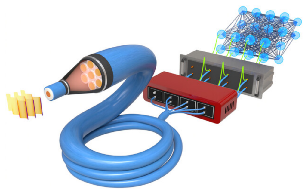
The sensor is built around a short segment of standard SMF28 fiber used as a few-mode waveguide at visible of near-infrared wavelengths. Light entering the fiber evolves into a stable speckle field whose structure depends sensitively on the input polarization. Instead of imaging this field, we sample only seven spots using a multimode fiber array that feeds single-photon avalanche diodes. Each polarization state produces a characteristic distribution of photon counts across the detectors. A deep neural network, trained on a set of accurately prepared polarization states, converts these seven numbers into the full polarization coherence matrix, allowing us to reconstruct both fully and partially polarized light.
Accuracy, speed, and stability
The sensor achieves an average infidelity around 10⁻³ on independently measured states, which is comparable to the accuracy of the polarization preparation used for calibration. It performs single-shot measurements at rates from a few tens to several thousand states per second depending on photon flux, and it operates comfortably at picowatt powers. A key practical result is extremely long stability. A 12 mm fiber segment fixed in a ceramic housing maintains accuracy for more than a month. Because intermodal coupling develops over a few hundred wavelengths, even shorter fibers could be used in the future.
Applications in imaging and dynamics
We tested the sensor on several representative samples. Scanning dense connective tissue reveals polarization textures unavailable from intensity images alone, with results matching those from a dedicated polarization microscope. Measurements on a freely drifting Actinoptychus heliopelta diatom show how the device captures birefringence patterns in moving, living specimens, something difficult to achieve with rotating-optic polarimeters. In a dynamic test, we track a voltage-induced polarization transition in a twisted-nematic liquid crystal with 5 ms temporal resolution, resolving its trajectory on the Poincaré sphere in real time.
Benchmarking and outlook
Comparison with a camera-based rotating-wave-plate polarimeter shows quantitative agreement with fidelities close to 0.99, and scans of a USAF birefringent target yield a spatial resolution of roughly 6 µm in the current geometry. The overall architecture illustrates how a random optical transform (optical front end) followed by a compact neural network (computational back end) can serve as an efficient optical sensor. The combination of single-photon sensitivity, speed, compactness, and long-term stability sets the stage for practical polarization-resolved microendoscopy and other applications where traditional polarimetry cannot be deployed.
